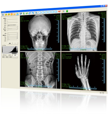
Easy Image Manipulation with NAOMI DICOM Imaging Software
NAOMI DICOM Imaging Software (Compatible with DICOM 3.0*) is the user-friendly operating software, increasing practicality for doctors and staffs to support smooth and effective clinical care. It enables you to adjust the images easily, enhancing its immense data from soft tissue to osseous parts with smoother and deeper contrast.
*DICOM (Digital Imaging and Communication in Medicine)
A communication standard developed by American College of Radiology (CAR) and National Electrical Manufacturers Association (NEMA) for a medical image format acquired from CT, MRI, or digital x-ray.
 Capture
 Print-Out
 Enhancement
 Negative-Positive Change Over
 Reflection
 Rotation
 Multi-Window Display
 Annotation/Measurement
 Setup
Setup
Easy Image Manipulation with a Single Click
on the Mouse
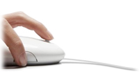
With NAOMI DICOM Imaging Software, all the helpful functions are available with a simple mouse operation. A single click does not only enable you to capture x-ray images, but also to easily zoom in and out on the images, adjusting brightness and contrast, and more with the designated icons on the software menu. DICOM image data also helps you to manage the patient’s information or date of x-rays, and sending images to remote locations. It is the absolutely user-friendly software for anybody.
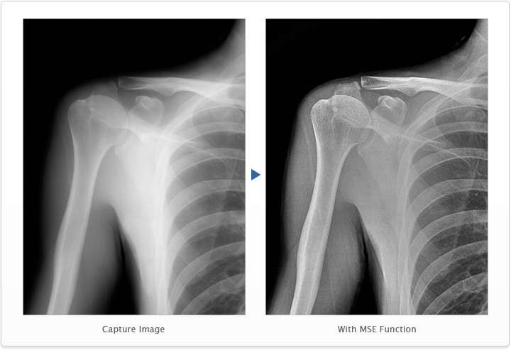
Enhancement Function
NAOMI DICOM Imaging Software displays the captured images in film-like softness or in a crisp view, achieved by the Multi-Scale Enhancement function. You can choose the image display modes depending on the application in order to provide a proper and more efficient diagnosis.
See more NAOMI sample images
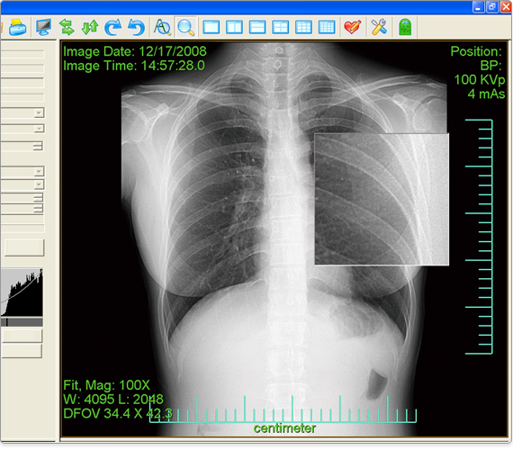
Zoom In & Out
You can zoom in and out on an area of interest for your diagnosis purpose or for better presentation for your patients.
Negative-Positive Change Over
By changing over its grayscale, it helps to find the area, which would have been difficult to see on a regular x-ray image.
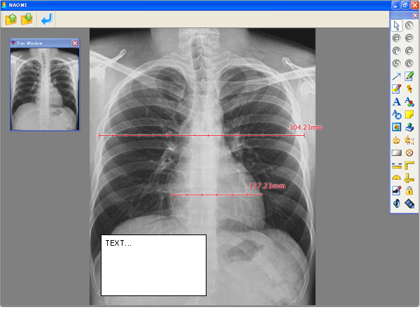
Annotation / Measurement
In Annotation Mode, you can measure the length and angle, or create and insert a comment in a text box, and save them with the image data.
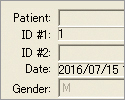
Patient Information
DICOM format saves the patient information and ID number with the image data file. (ID number is required to be entered.)
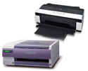
Printout
The captured images can be printed out with a printer or dry imager available in the market.